Module 2: Human Nervous System#
2.1 Introduction#
The Human nervous system consists of a brain, spinal cord, nerves and is one of the most complex and vital systems in the body, responsible for receiving, transmitting, and processing information. It acts as the body’s command center and enables communication between different parts of the body, allowing organisms to interact with their environment.
It is divided into two major parts:
Central Nervous System (CNS)
Peripheral Nervous System (PNS)
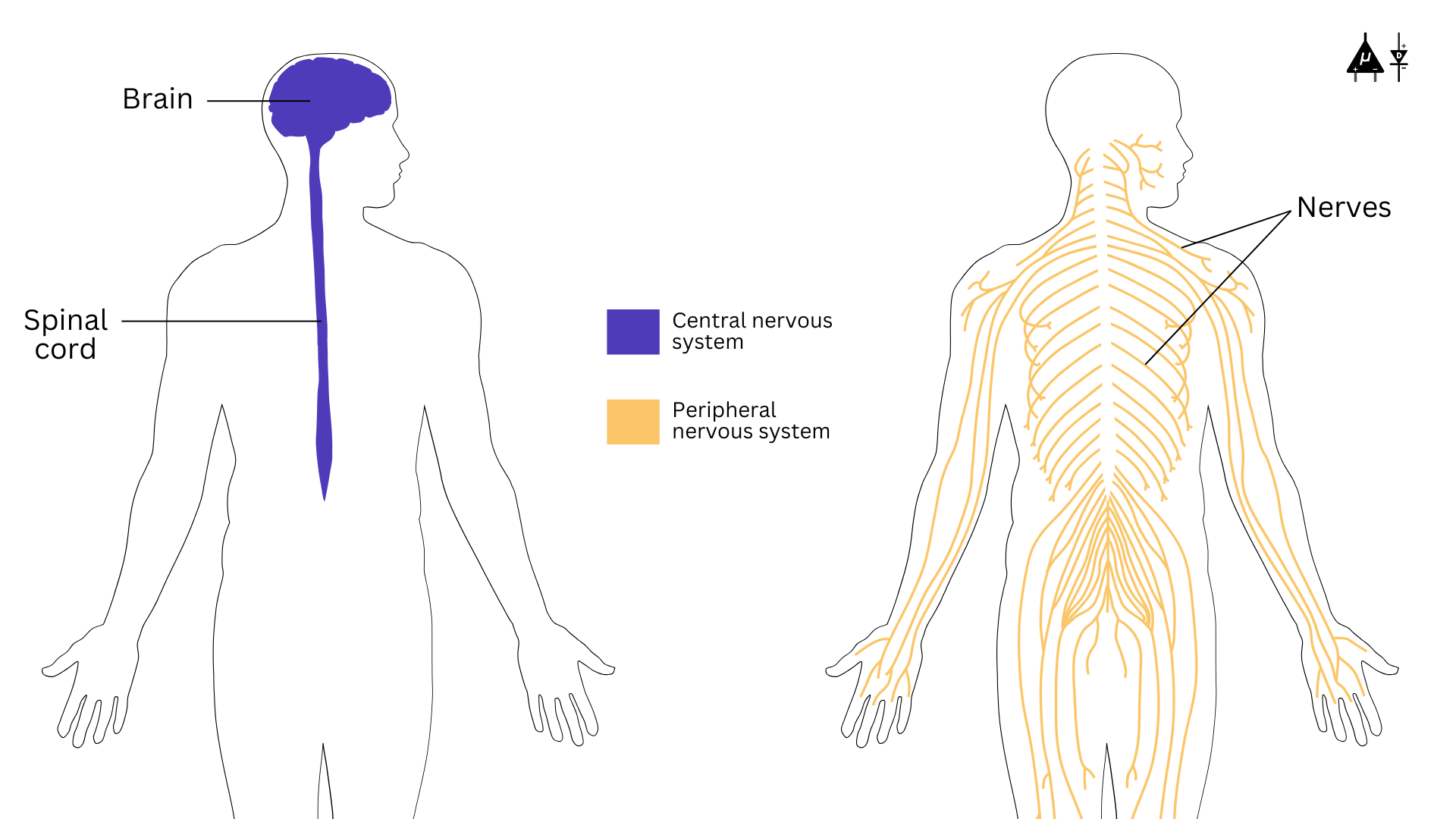
Types of nervous system#

An overview of the nervous system#
2.2 Central Nervous System (CNS)#
The central nervous system (CNS) is the body’s command center and is made up of your brain and spinal cord. The brain is protected by the cranium (also known as skull) while your vertebrae protects your spinal cord.
2.2.1 The Brain#
The brain is the most complex organ which communicates with the body by sending and receiving chemical and electrical signals. Some signals remain within the brain, while others are transmitted through the spinal cord and across a network of nerves to distant parts of the body. This communication relies on billions of neurons that form the central nervous system. Structure of the brain can be divided into 3 major parts.
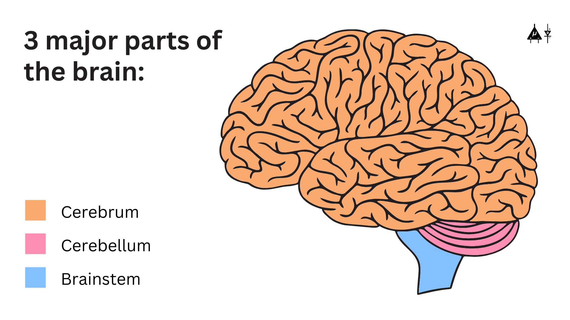
3 major parts of human brain#
Fun Fact
Brain weighs about 3 pounds in the average adult and contains about 60% fat. The remaining 40% is a combination of water, protein, carbohydrates and salts.
The brain itself is not a muscle. It contains blood vessels and nerves, including neurons and glial cells.
Cerebrum#
Cerebrum is the largest part of the brain which can be divided into 2 hemispheres, right and left.
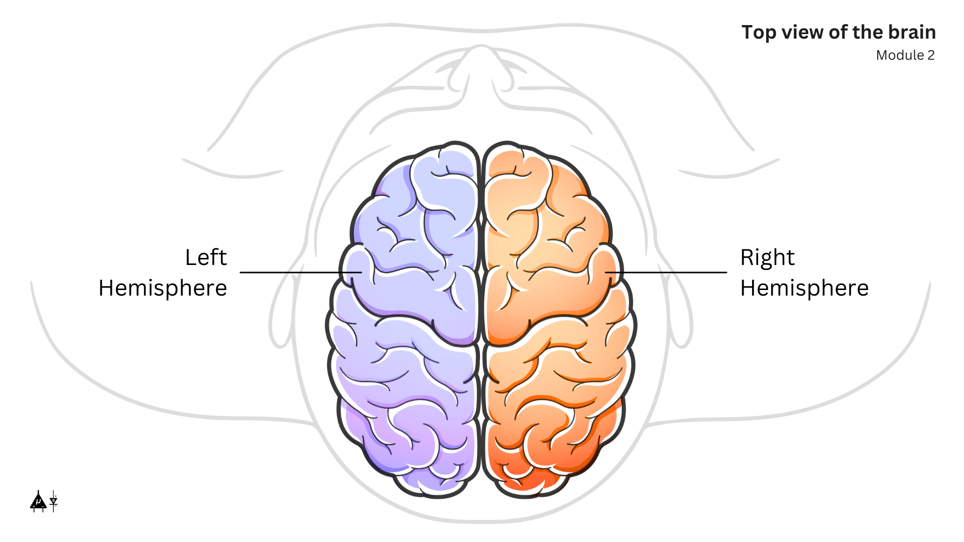
The 2 hemispheres of the brain#
Fun fact
The right hemisphere controls the left side of the body, and the left hemisphere controls the right side of the body.
The two halves communicate with one another through a large C shaped structure called corpus callosum which is the center part of the cerebrum.
Each hemisphere is further divided into four lobes:
Frontal lobe: The largest brain lobe, situated at the front of the head, the frontal lobe is involved in personality characteristics, decision-making, movement, speech and smell.
Parietal lobe: Located in the middle part of the brain, the parietal lobe helps a person identify objects and understand spatial relationships (where one’s body is compared with objects around the person). The parietal lobe is also involved in processing sensory information (touch, pain, temperature) and understanding spoken language.
Temporal lobe: Positioned on the sides of the brain, the temporal lobes are involved in short-term memory, speech, musical rhythm and some degree of smell recognition.
Occipital lobe: Found at the back of the brain, the occipital lobe is primarily responsible for processing visual information.
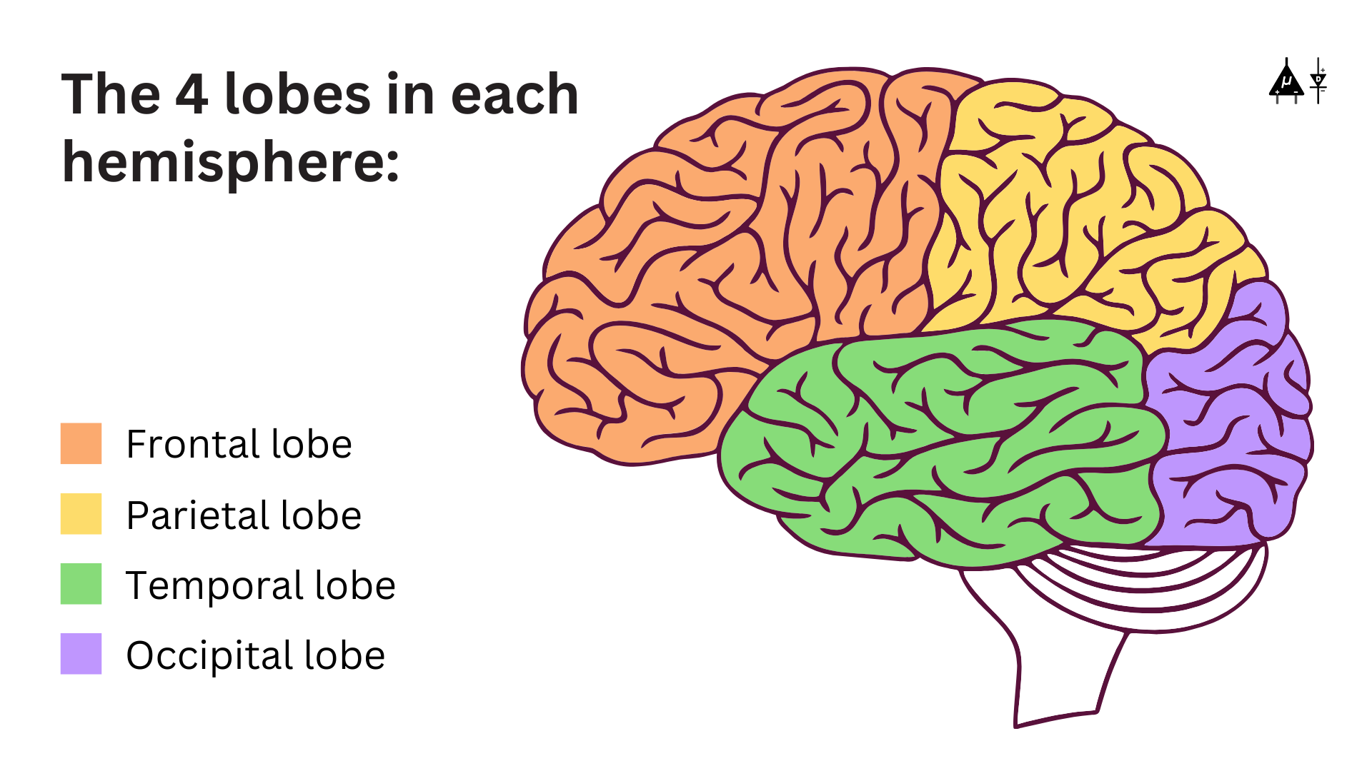
Different lobes of the brain#
Cerebellum#
The cerebellum (little brain) is a fist-sized portion of the brain located at the back of the head and above the brainstem. Its function is to coordinate voluntary muscle movements and to maintain posture, balance and equilibrium.
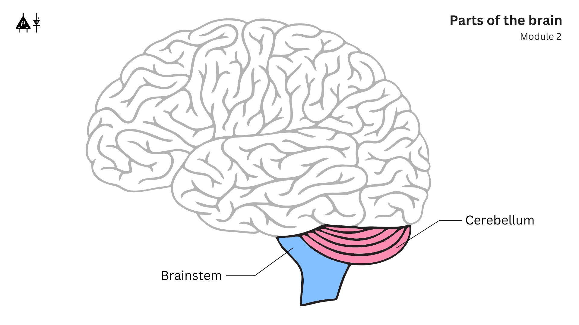
The cerebellum and brainstem#
Brainstem#
The brainstem (middle of brain) connects the cerebrum to the spinal cord. The brainstem includes the midbrain, the pons and the medulla.
Midbrain: Involved in motor control and auditory/visual processing.
Pons: It is a connection between midbrain and medulla. It controls sleep, respiration, and some motor functions.
Medulla: At the bottom of the brainstem, the medulla is where the brain meets the spinal cord. The medulla is crucial for survival, as it regulates vital bodily functions, including heart rate, breathing, blood circulation, and the balance of oxygen and carbon dioxide levels. It also controls reflexive actions such as sneezing, vomiting, coughing, and swallowing.
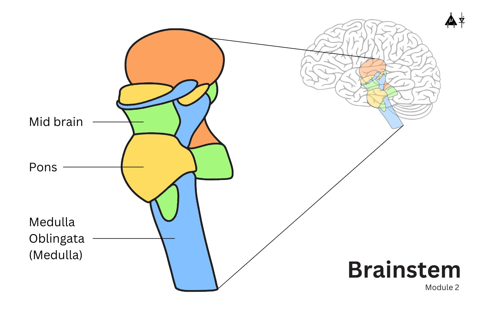
Overview of the brainstem#
2.2.2 The Spinal Cord#
The spinal cord begins at the base of the medulla and passes through a large opening at the bottom of the skull. Supported by the vertebrae, it serves as a communication highway between the brain and the rest of the body. This long, tubular structure transmits sensory information from the body to the brain and sends motor commands from the brain to the body. Additionally, it is responsible for reflex actions, which are quick and involuntary responses to stimuli.
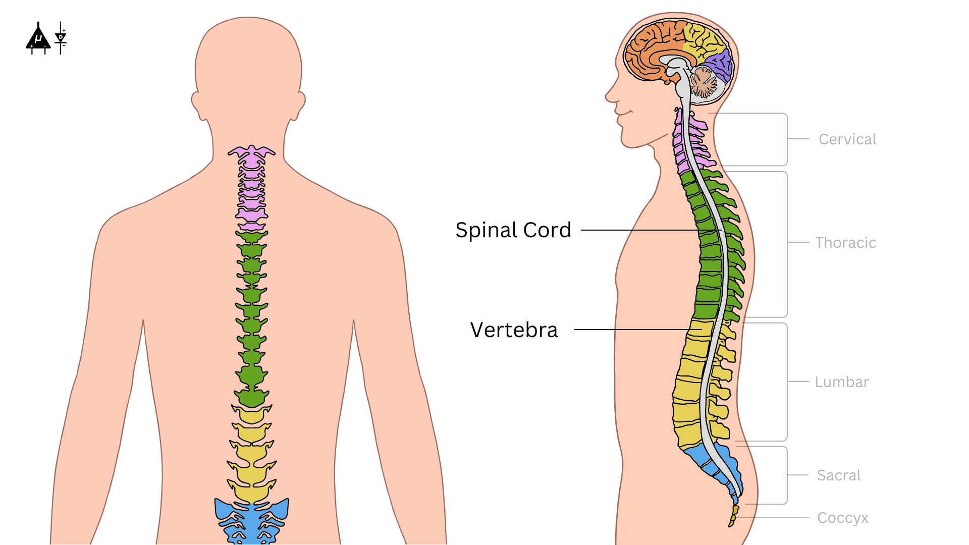
Spinal cord and vertebrae#
2.3 Peripheral Nervous System (PNS)#
The Peripheral Nervous System connects the Central Nervous System to the rest of the body and is responsible for transmitting signals to and from various organs and tissues. It is divided into two major systems:
2.3.1 Somatic Nervous System (SNS)#
The somatic nervous system controls voluntary movements and transmits sensory information to and from the central nervous system. It consists:
Sensory Neurons (Afferent Neurons): These neurons carry signals from sensory receptors (skin, muscles, joints) to the CNS, allowing us to perceive sensations like pain, temperature, and touch.
Motor Neurons (Efferent Neurons): These neurons transmit commands from the CNS to the skeletal muscles, enabling voluntary movement such as walking, talking, and picking up objects.
2.3.2 Autonomic Nervous System (ANS)#
The autonomic nervous system controls involuntary physiological processes, such as heart rate, digestion, and respiratory rate. It operates without conscious control and is divided into two main parts:
Sympathetic Nervous System: Known as the “fight or flight” system, it prepares the body for stress or emergency situations by increasing heart rate, dilating pupils, releasing adrenaline, and redirecting blood flow to muscles.
Parasympathetic Nervous System: It does the opposite of the sympathetic nervous system. Often referred to as the “rest and digest” system, it promotes relaxation by slowing the heart rate, promoting digestion, and conserving energy after a stressful event.
2.4 Neurons#
Neurons are the building blocks of the nervous system and are responsible for receiving and transmitting electrochemical signals throughout the body.
Fun Fact
Our brain is made up of about 80 billion neurons (that is 80,000,000,000).
2.4.1 Types of neurons#
Sensory Neurons: Transmit sensory information (e.g., pain, temperature, pressure) from receptors to the CNS.
Motor Neurons: Carry commands from the CNS to muscles and glands, enabling actions like muscle contraction or hormone release. It is the most common type of neuron.
Interneurons: These neurons are found in the CNS and act as connectors between sensory and motor neurons. They help process and integrate information.
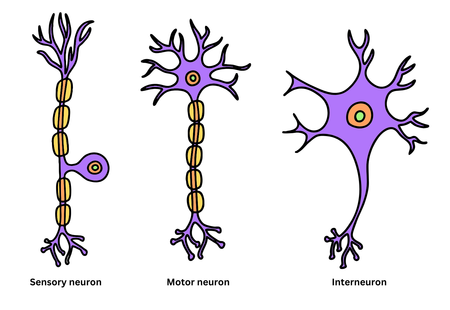
2.4.2 Structure of neuron#
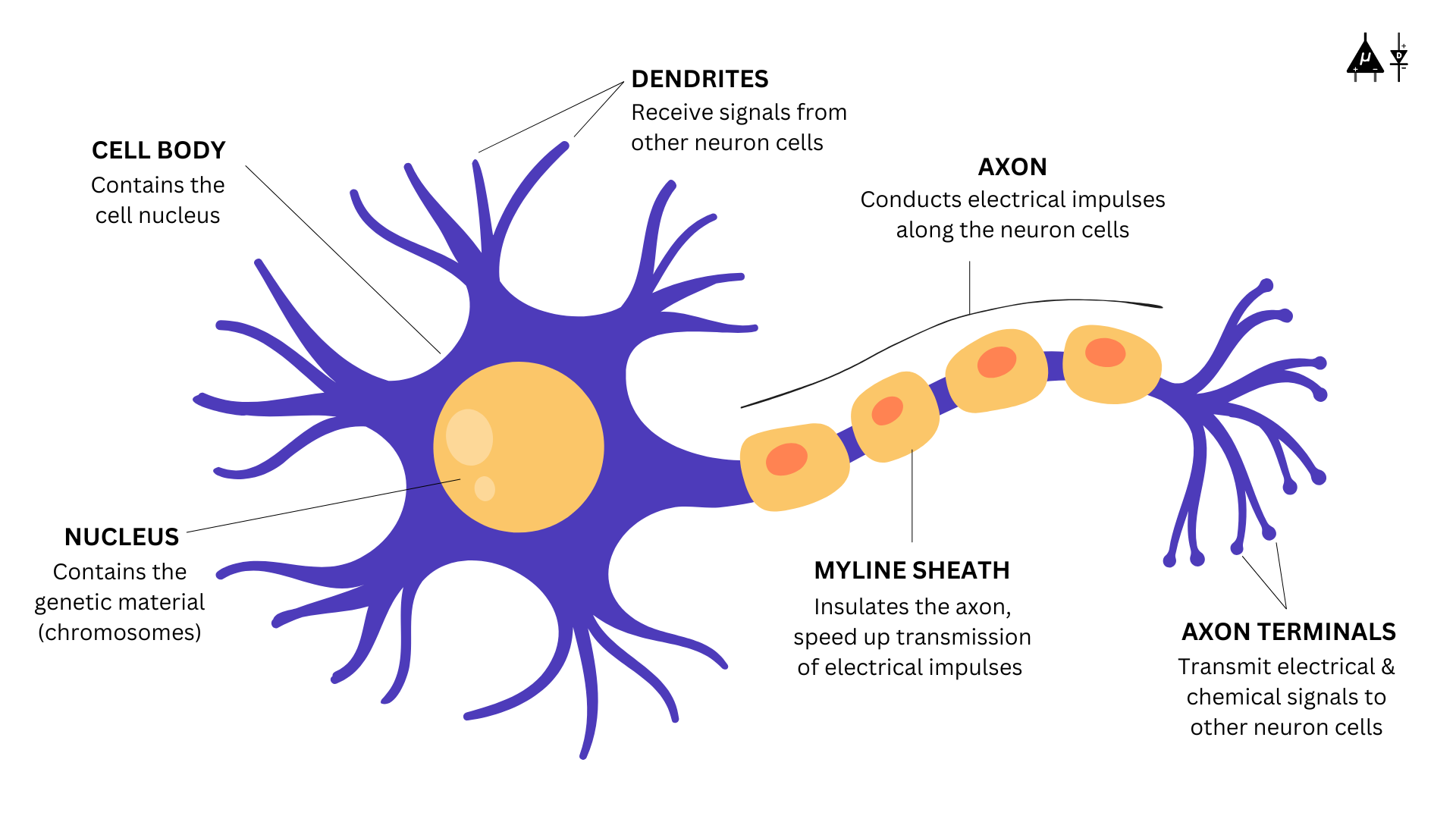
Structure of a neuron#
Cell Body (Soma): The soma, or cell body, is the core of the neuron which maintains the cell and to keep the neuron functioning efficiently. It is enclosed by a membrane that protects it and allows it to interact with its immediate surroundings
Nucleus: Nucleus contains the genetic material (chromosomes) of the neuron cell.
Dendrites: Dendrites are the tree root shaped part of the neuron which is responsible for receiving information from other neurons and to transmit electrical signals to the cell body.
Axons: Axons are the tail-like structure of the neuron which are responsible for transmitting electrical impulses (action potentials) away from the cell body toward other neurons.
Myelin sheath: Myelin sheath is a fatty layer that insulates the axon, speeding up signal transmission.
Synapse: Neurons do not touch each other, but where one neuron comes close to another neuron, a synapse is formed between the two which acts as a junction between two neurons where neurotransmitters are released to transmit signals to the next neuron.
Fun fact
There are axon-less neurons too where the signal is transmitted and received both by the dendrites.