Module 9 : EEG#
Basic Terminology#
Electroencephalography (EEG): Electroencephalography is a non-invasive technique used to record the electrical activity of the brain. It measures the electrical signals generated by neurons in the cerebral cortex using electrodes placed on the scalp.
Electroencephalograph: An electroencephalograph is the instrument used to record and amplify the brain’s electrical signals detected by scalp electrodes during electroencephalography.
Electroencephalogram: An electroencephalogram is the visual recording or waveform produced by the electroencephalograph, which represents the brain’s electrical activity over time.
Note
If you want to know detailed history of EEG, you can refer to this section: History of EEG
9.1 Introduction#
EEG is a non-invasive medical test that records the electrical activity of the brain using small electrodes placed on the scalp. It helps detect abnormal brain wave patterns and is commonly used to diagnose epilepsy, sleep disorders, brain injuries and for studying psychiatric conditions such as bipolar disorder and schizophrenia.

Normal EEG Frequency range : [1]
Delta waves: 0.5-4 Hz
Theta waves: 4-7 Hz
Alpha waves: 8-12 Hz
Beta waves: 13-30 Hz
Gamma waves: 30-80 Hz
9.2 Basic principle#
EEG measures the tiny electrical voltages (in microvolts) generated on the scalp when large groups of brain neurons fire synchronously. It mainly records the summed inhibitory and excitatory postsynaptic potentials from cortical pyramidal neurons, which get amplified and displayed as brain waves over time.
9.3 How it works#
9.3.1 Electrode Placement#
In surface EEG, small sensors called electrodes are placed on the scalp according to standardized systems like the 10-20 system, which ensures consistent positioning over brain regions.
Electrode Cap: A wearable cap that holds multiple electrodes in place. This ensures consistent electrode placement and is used for convenience during longer recordings.
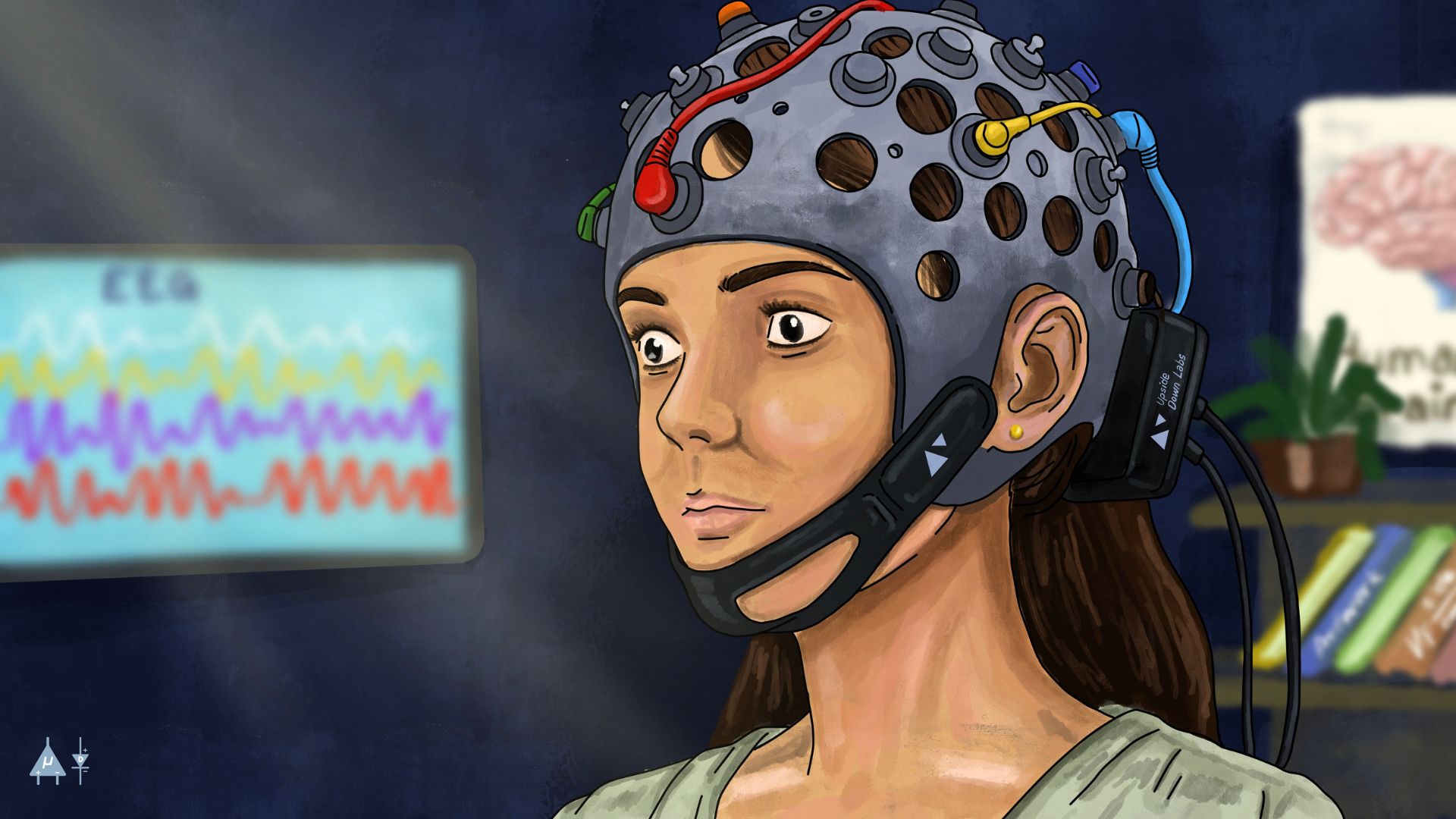
EEG electrode placement (Electrode cap)#
9.3.2 Signal Detection#
When groups of neurons in the brain fire synchronously, they generate tiny electrical fields that propagate to the scalp. The electrodes detect these voltage fluctuations, known as brain waves, which represent summed postsynaptic potentials from cortical neurons.
9.3.3 Signal Amplification and Filtering#
Since these signals are extremely weak (measured in microvolts), they are first amplified to make them readable. Then, unwanted noise, such as eye blinks, muscle artifacts, or 50/60 Hz power line interference-is filtered out to ensure the recorded data is clean and accurate.
Upside Down Labs Hardware compatible with EEG signal acquisition and processing:
We offer our own open source Chords software suite, featuring tools for signal visualization, data recording (with easy save and download options), time-based plotting, and a host of other benefits-such as analyzing signal frequencies and bandpower.
9.4 Key components of EEG#
9.4.1 Electrode placement system#
1. 10-20 system#
This is the standard system used for placing electrodes on the scalp. It divides the scalp into regions and assigns electrode positions based on specific anatomical landmarks (e.g., nasion, inion, and ear lobes). This system helps in the consistent placement of electrodes.
If you want to know the 10-20 system in detail , kindly refer to this section : 10-20 system wikipedia
2. High-Density EEG#
In some advanced setups, additional electrodes are placed for more detailed spatial information. This can involve 64, 128, or even 256 electrodes.
9.4.2 Characteristics of Different EEG Waves Types [1]#
Delta waves
Low frequency, high amplitude waves
Frequency range : 0.5 - 4 Hz
Associated with deep sleep (Non Rapid Eye Movement) stage 3 & 4 in adults
Amplitude is usually high (>75 microvolts) and can reach up to 200 microvolts in infants.
Delta waves are normally present during deep sleep and are also observed in infants, but they can be abnormal in awake adults, indicating brain injury or dysfunction.

Theta waves
Frequency range : 4-7 Hz
Associated with drowsiness and early sleep stages
Common during NREM stage 1 and 2 sleep, as well as during deep meditation or relaxation.
Also associated with creativity, memory processing and hippocampal activity, but excessive theta activity in awake adults can indicate neurological disorders or cognitive impairment.

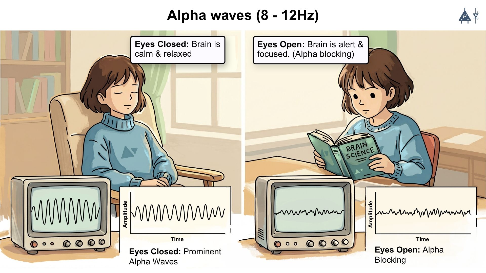
Alpha waves
Frequency range : 8-12 Hz
Associated with relaxed wakefulness, particularly when a person is awake but calm and not focused on specific tasks.
Alpha waves are most prominent when the eyes are closed.
Alpha waves decreases or disappears when a person opens their eyes, focuses attention, or engages in mental activity. This phenomenon is known as alpha blocking or alpha attenuation.
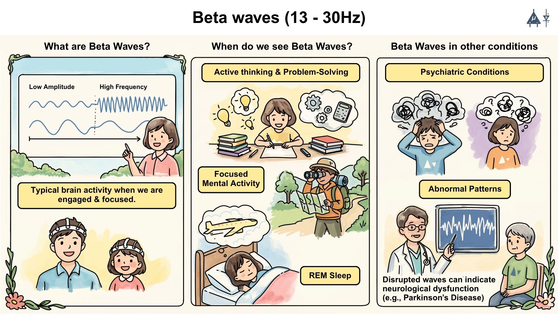
Beta waves
Frequency range : 13-30 Hz
Most common EEG pattern seen in awake, alert adults and children
Abnormal beta patterns can indicate neurological dysfunction such as in Parkinson’s disease.
These waves have relatively low amplitude and high frequency, and are associated with active thinking, problem-solving, and focused mental activity. They are also observed during REM sleep and in certain psychiatric conditions such as anxiety or schizophrenia.
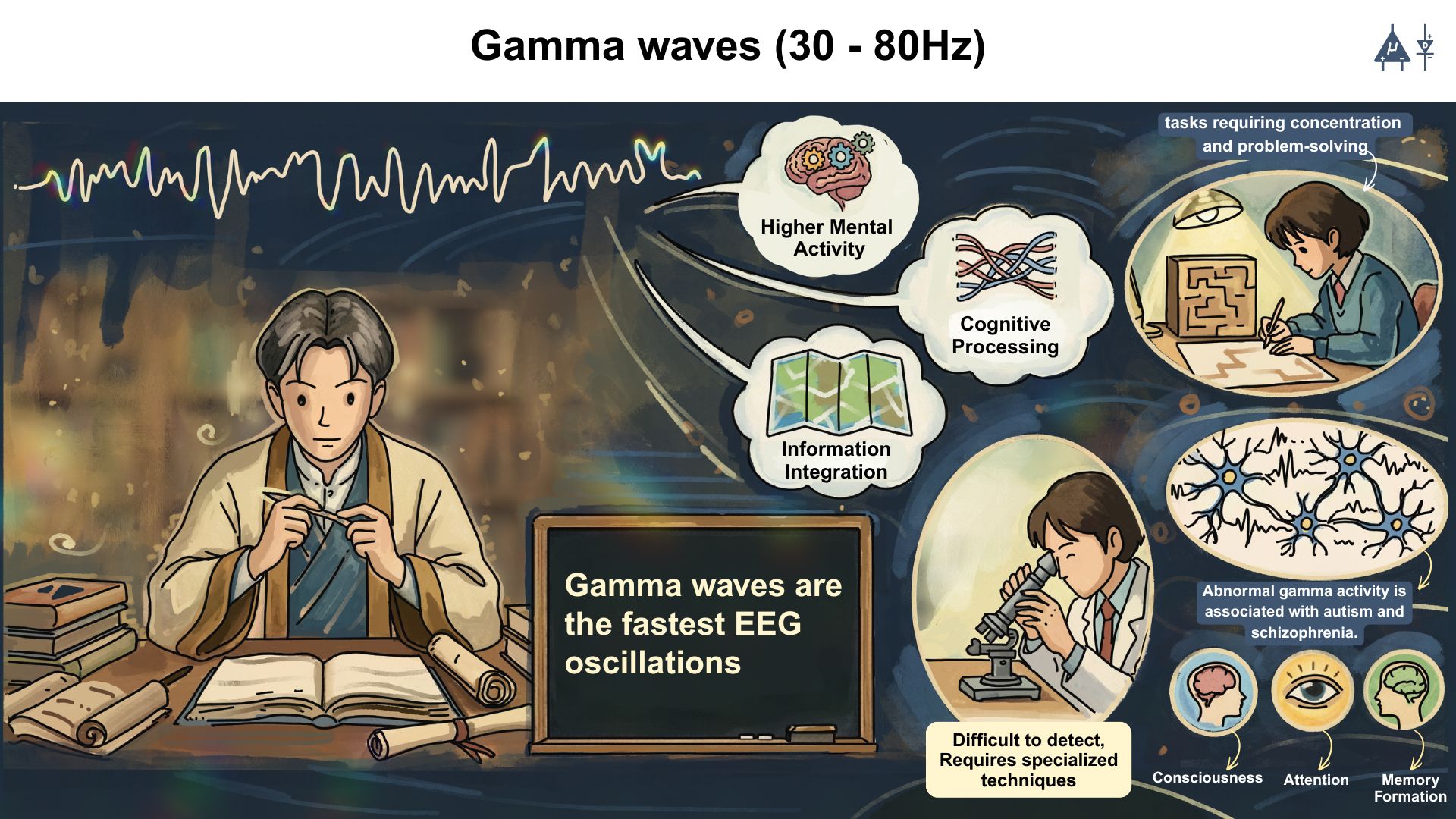
Gamma waves
Frequency range : 30-80 Hz
Associated with higher mental activity, cognitive processing, and information integration.
They are often seen in tasks requiring concentration and problem-solving.
Gamma waves are the fastest EEG oscillations.
They are thought to play a role in consciousness, attention, and memory formation. Abnormal gamma activity has been linked to conditions such as autism and schizophrenia.
Gamma waves are typically of low amplitude and can be difficult to detect with standard EEG equipment, often requiring specialized techniques for accurate measurement.
9.5 Applications of EEG#
1. Clinical Applications#
Diagnosis of Epilepsy: EEG is a key tool for diagnosing epilepsy. It can detect abnormal brain wave patterns, such as spikes or sharp waves, which indicate epileptic seizures. [2]
Sleep Disorders: EEG is used in sleep studies (polysomnography) to monitor sleep stages and identify disorders such as sleep apnea, insomnia, or narcolepsy. [3]
Monitoring of Brain Activity: EEG is used in critical care settings to monitor patients who are in coma, vegetative states, or under anesthesia. It can help assess brain function and guide treatment decisions.
Brain Death Diagnosis: EEG can be used to confirm brain death, which is characterized by the absence of electrical activity in the brain. [4]
Neurocognitive Disorders: EEG can help assess conditions such as dementia, Alzheimer’s disease, or encephalitis, as abnormal patterns of brain activity may be observed. [5]

EEG helps in diagnosis of brain disorders#
2. Research Applications#
Brain-Computer Interfaces (BCIs): EEG helps in BCIs by converting brain signals into commands that can control devices like artificial limbs or computers. This allows people who have difficulty moving to communicate or operate machines by using only their brain activity. [6]
Cognitive Neuroscience: EEG is widely used to study brain function during various cognitive tasks. Researchers use EEG to explore attention, memory, perception, and language processing. [7]

EEG applications in BCIs#
3. Neurofeedback#
EEG is also used in neurofeedback therapy, where individuals are trained to control their brain activity. This can be applied to treat conditions like ADHD, anxiety, depression, and even help with performance enhancement in athletes and musicians. [8]

EEG applications in neurofeedback therapy#
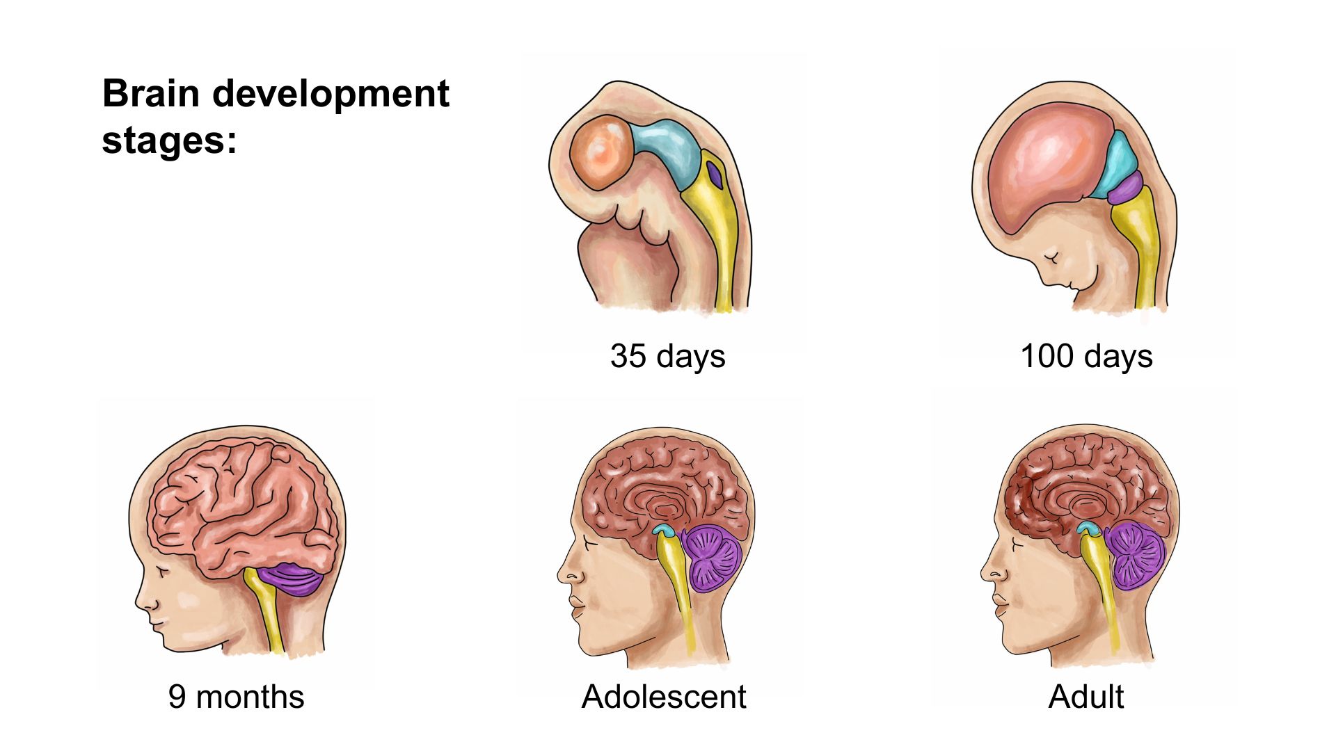
4. Brain Development and Aging#
EEG is used to investigate how brain activity changes across the lifespan. Researchers study EEG patterns in infants, children, adults, and elderly individuals to understand brain maturation, aging processes, and neurodevelopmental disorders.
5. Emotional and Affective Neuroscience#
EEG is used to study neural responses related to emotions, emotional regulation, and affective processing. Researchers examine brain oscillations and ERP components associated with emotional stimuli.
6. Attention and Cognitive Control Studies#
EEG is commonly used in experimental paradigms such as Stroop tasks, oddball tasks, and conflict monitoring tasks to study attention, cognitive control, and decision-making processes.
Projects Using EEG#
You can utilize our BioAmp Hardware to create various applications. We’ve successfully developed multiple applications, so there’s nothing holding you back from creating something innovative and outstanding. A few applications of our devices are highlighted below:
9.6 Advantages of EEG#
Non-invasive & safe
Cost-effective
Useful for diagnosing neurological disorders
Real-time monitoring
Suitable for long-duration monitoring
Can be used in cognitive and psychophysiological research
Portable & accessible
Direct measure of brain activity
9.7 Limitations of EEG#
Poor Spatial Resolution: EEG cannot provide detailed spatial information about the precise location of brain activity, as it measures only the electrical activity at the scalp surface.
Requires expert analysis: Interpretation can be complex and subjective.
Sensitivity to External Noise: EEG signals can be distorted by artifacts caused by muscle movements (such as eye blinks or facial contractions) and external electrical interference, which may mask the brain’s actual electrical activity.
Limited Depth Information: EEG is primarily sensitive to the activity of cortical neurons, and it is not as effective in detecting activity from deeper structures of the brain, such as the brainstem or subcortical areas.
Limited to Large Scale Potentials: EEG primarily records the summed electrical activity of large populations of neurons, especially cortical pyramidal cells. Activity from small or isolated neuronal groups is usually too weak to be detected on the scalp.
Inverse Problem: Determining the exact brain source of recorded scalp potentials is mathematically difficult. Multiple possible neural sources can produce similar EEG patterns.
Poor Signal-to-Noise Ratio (SNR): Brain signals recorded by EEG are very small (in microvolts) and are often mixed with noise, making it challenging to isolate meaningful neural activity.
9.8 Summary#
EEG is a non-invasive technique that records the brain’s electrical activity using scalp electrodes. It helps diagnose conditions such as epilepsy, sleep disorders, and psychiatric illnesses, and is also used in cognitive research and brain computer interfaces. Although it has limitations like poor spatial resolution and susceptibility to noise, EEG remains highly valuable due to its real-time monitoring capability and excellent temporal precision.
Warning
This content is provided solely for educational purposes. Always seek medical advice from a healthcare expert for clinical application.